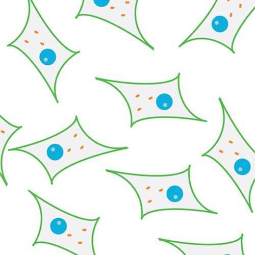
L6-GLUT4myc Rat Myoblast Cell Line
Immortalized rat skeletal muscle cells (L6) over-expresses GLUT4 with a myc-epitope, which is useful for measuring GLUT4 translocation.
Highlights:
- L6 myoblast cell line that stably expresses GLUT4 protein containing a fourteen amino acid epitope of human c-myc within its first exofacial loop
- Only cellular model available to study glucose uptake and GLUT4 translocation without permeabilizing or fractionating the cells
- Useful for novel anti-diabetic compound screening
- Amendable for transient transfection by plasmid-based gene transfer and viral infection protocols (retrovirus and adenovirus)
Insulin stimulation of glucose transport in skeletal muscle results mainly due to translocation of the glucose transporter GLUT4 to the cell surface. L6 cells, originally derived from rat skeletal muscle, propagate as mononucleated myoblasts but can differentiate into multinucleated primary myotubes. The myotubes express several proteins typical of skeletal muscle including the GLUT4 glucose transporter. Insulin stimulates glucose uptake with high sensitivity and maximal responsiveness only in differentiated L6 myotubes and GLUT4 expression parallels the acquisition of these characteristics as the L6 cells differentiate. These features of L6 myotubes are important since GLUT4 is responsible for insulin-dependent glucose uptake in mature skeletal muscle.
Also available: L6 Rat Myoblast Cell Line
From the laboratory of Amira Klip, PhD, Hospital For Sick Children.
This product is for sale to Nonprofit customers only. For profit customers, please Contact Us for more information.
| Product Type: | Cell Line |
| Name: | L6-GLUT4myc |
| Cell Type: | Skeletal muscle |
| Accession ID: | CVCL_0P25 |
| Morphology: | Myoblast/Myotube |
| Source: | Quadriceps |
| Organism: | Rat |
| Biosafety Level: | II |
| Growth Conditions: | MEM-α (with Ribonucleosides and Deoxyribonucleosides) +10% FBS, 1% Antimycotic +5 ug/ml blasticidin |
| Subculturing: | 2-3 days |
| Cryopreservation: | MEM-α +10% FBS+ 10% DMSO |
| Comments: | The myc-epitope in the GLUT4myc has the amino acid sequence of AEEQKLISEEDLLK |
| Storage: | Liquid nitrogen |
| Shipped: | Dry ice |
L6 myoblasts stably express rat GLUT4myc glucose transporter, a chimera of rat GLUT4 with human myc-epitope (AEEQKLISEEDLLK; recognized by monoclonal antibody clone 9E10) in the first exofacial loop of the transporter (between residue 66 and 67). Myoblasts should be passaged at approximately 70% of confluence. Myoblasts can be differentiated into myotubes by switching confluent myoblast monolayers to MEM-a +2% FBS for 4-5 days.*Antibiotic resistance for the GLUT4 stable transfection is blasticidin-HCl and passaging myoblasts with blasticidin in the growth media is recommended to maintain selection pressure.
Cell Line References
- Wang Q, Khayat Z, Kishi K, Ebina Y, Klip A. (1998) GLUT4 translocation by insulin in intact muscle cells: detection by a fast and quantitative assay. FEBS Letters 427: 193-7;
- Ueyama A, Yaworsky KL, Wang Q, Ebina Y, Klip A. (1999) GLUT-4myc ectopic expression in L6 myoblasts generates a GLUT-4-specific pool conferring insulin sensitivity. Am. J. Physiol. Endocrinol. Metab. 277: E572-8;
- Ishikura S, Antonescu CN, Klip A. (2010) Documenting GLUT4 Exocytosis and Endocytosis in Muscle Cell Monolayers. Curr. Protoc. Cell Biol., John Wiley & Sons, Inc., 46: Unit 15.15: 1-9;
- Rudich A, Konrad D, Török D, Ben-Romano R, Huang C, Niu W, Garg RR, Wijesekara N, Germinario RJ, Bilan PJ, Klip A. (2003)Indinavir uncovers different contributions of GLUT4 and GLUT1 towards glucose uptake in muscle and fat cells and tissues. Diabetologia 46: 649-58.
- Huang C, Somwar R, Patel N, Niu W, Török D, Klip A. Sustained exposure of L6 myotubes to high glucose and insulin decreases insulin-stimulated GLUT4 translocation but upregulates GLUT4 activity. Diabetes. 2002 Jul;51(7):2090-8.
Application References
- Pillon NJ, Li YE, Fink LN, Brozinick JT, Nikolayev A, Kuo MS, Bilan PJ, KlipA. (2014) Nucleotides released from palmitate-challenged muscle cells through pannexin-3 attract monocytes. Diabetes. 63: 3815-26.
- Li Q, Zhu X, Ishikura S, Zhang D, Gao J, Sun Y, Contreras-Ferrat A, Foley KP, Lavandero S, Yao Z, Bilan PJ, Klip A, Niu W. (2014) Ca²? signals promote GLUT4 exocytosis and reduce its endocytosis in muscle cells. Am J Physiol Endocrinol Metab. Jul 15;307(2):E209-24.
- Sun Y, Chiu TT, Foley KP, Bilan PJ, Klip A. (2014) Myosin Va mediates Rab8A-regulated GLUT4 vesicle exocytosis in insulin-stimulated muscle cells. Mol Biol Cell. 25: 1159-70.
- Foley KP, Klip A. (2014) Dynamic GLUT4 sorting through a syntaxin-6 compartment in muscle cells is derailed by insulin resistance-causing ceramide. Biol Open. 3: 314-25.
- Chiu TT, Sun Y, Koshkina A, Klip A. (2013) Rac-1 superactivation triggers insulin-independent glucose transporter 4 (GLUT4) translocation that bypasses signaling defects exerted by c-Jun N-terminal kinase (JNK)- and ceramide-induced insulin resistance. J Biol Chem. 288:17520-31.
- Pillon NJ, Arane K, Bilan PJ, Chiu TT, Klip A. (2012) Muscle cells challenged with saturated fatty acids mount an autonomous inflammatory response that activates macrophages. Cell Commun Signal. 10: 30.
- Boguslavsky S, Chiu T, Foley KP, Osorio-Fuentealba C, Antonescu CN, Bayer KU, Bilan PJ, Klip A. (2012) Myo1c binding to submembrane actin mediates insulin-induced tethering of GLUT4 vesicles. Mol Biol Cell. 23: 4065-78.
- Kewalramani G, Fink LN, Asadi F, Klip A. (2011) Palmitate-activated macrophages confer insulin resistance to muscle cells by a mechanism involving protein kinase C ? and ?. PLoS One. 6: e26947.
- Sun Y, Bilan PJ, Liu Z, Klip A. (2010) Rab8A and Rab13 are activated by insulin and regulate GLUT4 translocation in muscle cells. Proc Natl Acad Sci U S A. 107: 19909-14.
- Ishikura S, Antonescu CN, Klip A. (2010) Documenting GLUT4 Exocytosis and Endocytosis in Muscle Cell Monolayers. Curr. Protoc. Cell Biol., John Wiley & Sons, Inc., 46: Unit 15.15: 1-9.
- Samokhvalov V, Bilan PJ, Schertzer JD, Antonescu CN, Klip A. (2009) Palmitate- and lipopolysaccharide-activated macrophages evoke contrasting insulin responses in muscle cells. Am J Physiol Endocrinol Metab. 296: E37-46.
- Antonescu CN, Díaz M, Femia G, Planas JV, Klip A. (2008) Clathrin-dependent and independent endocytosis of glucose transporter 4 (GLUT4) in myoblasts: regulation by mitochondrial uncoupling. Traffic. 9: 1173-90.
- Thong FS, Bilan PJ, Klip A. (2007) The Rab GTPase-activating protein AS160 integrates Akt, protein kinase C, and AMP-activated protein kinase signals regulating GLUT4 traffic. Diabetes. 56: 414-23.
- Wijesekara N, Tung A, Thong F, Klip A. (2006) Muscle cell depolarization induces again in surface GLUT4 via reduced endocytosis independently of AMPK. Am J Physiol Endocrinol Metab. 290: E1276-86.
- Ishiki M, Randhawa VK, Poon V, Jebailey L, Klip A. (2005) Insulin regulates the membrane arrival, fusion, and C-terminal unmasking of glucose transporter-4 via distinct phosphoinositides. J Biol Chem. 280: 28792-802.
- Rudich A, Konrad D, Török D, Ben-Romano R, Huang C, Niu W, Garg RR, Wijesekara N, Germinario RJ, Bilan PJ, Klip A. (2003) Indinavir uncovers different contributions of GLUT4 and GLUT1 towards glucose uptake in muscle and fat cells and tissues. Diabetologia 46: 649-58.
- Wang Q, Somwar R, Bilan PJ, Liu Z, Jin J, Woodgett JR, Klip A. (1999) Protein kinase B/Akt participates in GLUT4 translocation by insulin in L6 myoblasts. Mol Cell Biol. 19: 4008-18.
- Wang Q, Khayat Z, Kishi K, Ebina Y, Klip A. (1998) GLUT4 translocation by insulin in intact muscle cells: detection by a fast and quantitative assay. FEBS Letters 427: 193-7.
- Gao MH, Giamouridis D, Lai NC, Walenta E, Paschoal VA, Kim YC, Miyanohara A, Guo T, Liao M, Liu L, Tan Z, Ciaraldi TP, Schenk S, Bhargava A, Oh DY, Hammond HK. One-time injection of AAV8 encoding urocortin 2 provides long-term resolution of insulin resistance. JCI Insight. 2016 Sep 22;1(15):e88322. View Article
- Tunduguru R, Zhang J, Aslamy A, Salunkhe VA, Brozinick JT, Elmendorf JS, Thurmond DC. The actin-related p41ARC subunit contributes to p21-activated kinase-1 (PAK1)-mediated glucose uptake into skeletal muscle cells. J Biol Chem. 2017 Sep 25. pii: jbc.M117.801340. View Article
- Yang WM, Min KH, Son YS, Park SW, Lee W. Data on the expression and insulin-stimulated phosphorylation of IRS-1 by miR-96 in L6-GLUT4myc myocytes. Data Brief. 2017 Oct 28;15:728-732. doi: 10.1016/j.dib.2017.10.054. eCollection 2017 Dec. View Article
- Kellogg DL 3rd, McCammon KM, Hinchee-Rodriguez KS, Adamo ML, Roman LJ. Neuronal nitric oxide synthase mediates insulin- and oxidative stress-induced glucose uptake in skeletal muscle myotubes. Free Radic Biol Med. 2017 Sep;110:261-269. View Article
- Stierwalt HD, Ehrlicher SE, Bergman BC, Robinson MM, Newsom SA. Insulin-stimulated Rac1-GTP binding is not impaired by palmitate treatment in L6 myotubes. Physiol Rep. 2018 Dec;6(24):e13956. View Article
- Azhar S, Bittner S, Hu J, Shen WJ, Cortez Y, Hao X, Han L, Lagerstedt JO, Kraemer FB, Johansson JO. Novel ABCA1 peptide agonists with antidiabetic action. Mol Cell Endocrinol. 2019 Jan 15;480:1-11. View Article
- Min KH, Yang WM, Lee W. Saturated fatty acids-induced miR-424-5p aggravates insulin resistance via targeting insulin receptor in hepatocytes. Biochem Biophys Res Commun. 2018 Sep 10;503(3):1587-1593. View Article
- Jaldin-Fincati JR, Bilan PJ, Klip A. GLUT4 Translocation in Single Muscle Cells in Culture: Epitope Detection by Immunofluorescence. Methods Mol Biol. 2018;1713:175-192. View Article
- Zhang J, Oh E, Merz KE, Aslamy A, Veluthakal R, Salunkhe VA, Ahn M, Tunduguru R, Thurmond DC. DOC2B promotes insulin sensitivity in mice via a novel KLC1-dependent mechanism in skeletal muscle. Diabetologia. 2019 May;62(5):845-859. doi: 10.1007/s00125-019-4824-2. Epub 2019 Feb 1. View Article
- Bone DBJ, Meister J, Knudsen JR, Dattaroy D, Cohen A, Lee R, Lu H, Metzger D, Jensen TE, Wess J. Skeletal Muscle-Specific Activation of G(q) Signaling Maintains Glucose Homeostasis. Diabetes. 2019 Jun;68(6):1341-1352. View Article
- Kalinovich A, Dehvari N, Åslund A, et al. Treatment with a β-2-adrenoceptor agonist stimulates glucose uptake in skeletal muscle and improves glucose homeostasis, insulin resistance and hepatic steatosis in mice with diet-induced obesity. Diabetologia. 2020;63(8):1603-1615. View Article
- Min KH, Lee W. Alteration of mitochondrial DNA content modulates antioxidant enzyme expressions and oxidative stress in myoblasts. Korean J Physiol Pharmacol. 2019 Nov;23(6):519-528. View Article
- Fujimoto BA, Young M, Carter L, Pang APS, Corley MJ, Fogelgren B, Polgar N. The exocyst complex regulates insulin-stimulated glucose uptake of skeletal muscle cells. Am J Physiol Endocrinol Metab. 2019 Dec 1;317(6):E957-E972. View Article
- Masson SWC, Sorrenson B, Shepherd PR, Merry TL. β-catenin regulates muscle glucose transport via actin remodelling and M-cadherin binding. Mol Metab. 2020 Dec;42:101091. View Article
If you publish research with this product, please let us know so we can cite your paper.


