Optimized ECL Reagent
Buffered ECL reagent with optimized pH, provides a stronger and more stable chemiluminescent signal for western blots, compared to other commercial ECL reagents.
Highlights:
- Increased sensitivity and signal, compared to other commercial ECL reagents
- Faster exposure times (10-20 sec, compared to 2+ min)
- Increased signal stability over time (~3hrs)
Western blot is a widely used analytical technique to detect specific proteins from a complex mixture of proteins extracted from cells or tissue. The technique uses three elements: (1) separation by size, (2) transfer to a solid support, and (3) marking the target protein with specific primary and secondary antibody to visualize. The most popular method used to visualize a protein at the nanogram level involves enhanced chemiluminescence or ECL. The chemiluminescent (CL) reaction of luminol (the active ingredient in the buffer) is catalyzed by superoxide anions which are produced emzymatically from H202. The superoxides then convert the luminol to aminothalate ions, which gives rise to the chemiluminescence light at 430nm for visual detection of your protein.
From the laboratory of Michael A. Moxley, PhD and Abdul Waheed, PhD, St. Louis University.
 Part of The Investigator's Annexe program.
Buffered ECL reagent with optimized pH, provides a stronger and more stable chemiluminescent signal for western blots, compared to other commercial ECL reagents.
Highlights:
- Increased sensitivity and signal, compared to other commercial ECL reagents
- Faster exposure times (10-20 sec, compared to 2+ min)
- Increased signal stability over time (~3hrs)
Western blot is a widely used analytical technique to detect specific proteins from a complex mixture of proteins extracted from cells or tissue. The technique uses three elements: (1) separation by size, (2) transfer to a solid support, and (3) marking the target protein with specific primary and secondary antibody to visualize. The most popular method used to visualize a protein at the nanogram level involves enhanced chemiluminescence or ECL. The chemiluminescent (CL) reaction of luminol (the active ingredient in the buffer) is catalyzed by superoxide anions which are produced emzymatically from H202. The superoxides then convert the luminol to aminothalate ions, which gives rise to the chemiluminescence light at 430nm for visual detection of your protein.
From the laboratory of Michael A. Moxley, PhD and Abdul Waheed, PhD, St. Louis University.
 Part of The Investigator's Annexe program.
| Product Type: | Buffer or Chemical |
| Name: | Optimized ECL Reagent |
| Recommended Membrane: | PVDF |
| Components: | Luminol Stock Solution 250uL, Luminol Buffer 250mL |
| Recommended Film: | Hyperfilm ECL |
| Tested Applications: | Western Blot |
| Storage: | Luminol Stock: Keep dark at room temperature; Luminol Buffer: store at 4C |
| Shipped: | Cold packs |
Western Blot

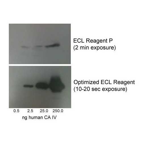
Western Blot with increasing amounts of CA IV protein. (top) Competitor ECL Reagent, 2 min film exposure. (bottom) Optimized ECL Reagent, 10-20 sec exposure. Primary antibody used: rabbit anti-human CA IV (1:5000); Secondary antibody used: goat anti-rabbit IgG peroxidase (1:10000).
Adapted from: Khan P, et al. Appl Biochem Biotechnol. 2014 May;173(2):333-55
Protocol Notes: Incubate (PVDF) membrane with 5 mL (for mini gel) or 15 mL (for large gel) of Luminol buffer containing 5 or 15uL, respectively of Luminol stock solution at room temperature for 5 min. Follow with exposure using x-ray film in dark room or image with CCD camera.
With exception to the above, it is otherwise recommended to follow normal Western Blotting procedures
- Khan P, Idrees D, Moxley MA, Corbett JA, Ahmad F, von Figura G, Sly WS, Waheed A, Hassan MI. Luminol-based chemiluminescent signals: clinical and non-clinical application and future uses. Appl Biochem Biotechnol. 2014 May;173(2):333-55.
If you publish research with this product, please let us know so we can cite your paper.


