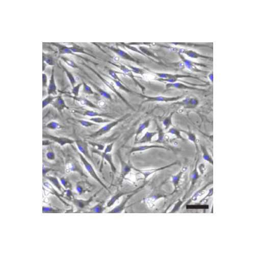
Rat Epineurial Cells
Primary cells isolated from the epineurium layer of the adult rat sciatic nerve.
Highlights:
- Cells are identified as S100 negative, GFAP negative, p75NGFR negative cells with a variable proportion of Thy-1 positive fibroblasts
- Highly proliferative and expandable for direct use or cryopreservation
- Thawing and re-plating of epineurial cells does not alter their viability or other phenotypic characteristics of the cells
- Cells can be used to study their effects on nerve regeneration
- They can be used as negative or reference controls in Schwann cell studies performed in vitro and in vivo
Primary cultures of epineurial cells can be used in a variety of applications, as it is becoming increasingly clear that these cells may have a beneficial role in nerve regeneration. Epineurial cells can be used along with Schwann cells to test the cell type specificity of the cellular responses in experiments carried out in vitro and in vivo. The availability of highly viable, cryopreserved stocks of epineurial cells can enable investigators to use the cells themselves or the products derived therefrom (such as DNA/RNA, proteins, exosomes, other) directly in scientific research. Due to their high proliferation rate and expandability, the cells have potential for continued culture, cryopreservation and used as platforms for drug or genetic screens.
From the laboratory of Paula V. Monje, PhD, University of Miami.
| Product Type: | Cell Line |
| Cell Type: | Primary epineurial cells from adult rat sciatic nerve tissue |
| Morphology: | Typical fibroblastic morphology, expanded, adherent |
| Source: | Sciatic nerve epineurium |
| Organism: | Rat - 3 months old female |
| Biosafety Level: | BSL-1 |
| Subculturing: | 2-3 additional passages (recommended). Extensive passaging is possible but the effect on phenotypic stability has not been tested |
| Growth Conditions: | DMEM high glucose containing 10 % decomplemented fetal bovine serum (FBS) and GlutamaxTM. Addition of antibiotics (penicillin, streptomycin, and gentamicin) does not impair viability. |
| Cryopreservation: | RecoveryTM or 90% FBS containing 10% DMSO |
| Comments: |
- Exhibit typical morphological features of cultured fibroblasts and high proliferative capicity. Some heterogeneity in both the shape and size of individual cells may be apparent. - Cells have a tendency to migrate and spontaneously form aggregates that resemble spheroids when plated at high densities or after reaching confluency. The effect of extended passaging on these populations has not been tested. Other properties of the cells have not yet been investigated. |
| Storage: | Liquid nitrogen |
| Shipped: | Dry Ice |
Microscopy Image

Cultures of adult epineurial cells from rat sciatic nerve. Upper panels: representative cultures immunostained with antibodies against Thy-1 (left panel, low magnification image) or Thy-1 and S100 (right panel, high magnification image)to reveal the presence of fibroblasts (Thy-1 positive, green) and absence of contaminating Schwann cells (S100 positive, red). Lower panels: high magnification view of a culture stained with Thy-1 antibodies (green) and EdU (which labels proliferating cells, red). Blue channel: DAPI (nuclear stain). Bottom right panel: phase contrast microscopy of a representative culture 3 days after plating.
Adapted from: Andersen et al. Scientific Reports 6, 31781.
- Andersen N, Srinivas S, Piñero G, Monje P.V. (2016) A rapid and versatile method for the isolation, purification and cryogenic storage of Schwann cells from adult rodent nerves. Scientific Reports 6, 31781; doi: 10.1038/srep31781.
If you publish research with this product, please let us know so we can cite your paper.


