Goat Anti-Doublecortin / DCX (aa232-242) Antibody
This goat IgG polyclonal antibody was generated against peptide sequence CKTSANMKAPQS from the internal region of neuronal migration protein doublecortin (DCX) and recognizes human, mouse, and rat DCX.
Highlights:
- Reacts with human, mouse, and rat DCX
- Suitable for Peptide ELISA, Western Blot, Immunohistochemistry, and Immunofluorescence applications
- Mutations in the DCX gene result in lissencephaly or subcortical band heterotopia
Neuronal migration protein doublecortin (DCX) is a microtubule-associated protein that is involved in neuronal migration in embryonic and postnatal brain development. DCX regulates the stability and organization of microtubules by influencing microtuble rigidity, curvature, and the 13-protofilament geometry. Mutations in the DCX gene can lead to epilepsy, cognitive disability, subcortical band heterotopia, and lissencephaly.
| Product Type: | Antibody |
| Name: | Goat Anti-Doublecortin / DCX (aa232-242) Antibody |
| Alternative Name(s): | Doublin, Lissencephalin-X (Lis-X) |
| Accession ID: | NP_000546.2; NP_835365.1; NP_835364.1; NP_001182482.1 |
| Antigen: | Doublecortin / DCX (aa232-242) |
| Isotype: | IgG |
| Clonality: | Polyclonal |
| Reactivity: | Human, Mouse, Rat |
| Specificity: | Doublecortin / DCX (aa232-242) |
| Immunogen: | CKTSANMKAPQS |
| Species Immunized: | Goat |
| Epitope: | Internal Region |
| Purification Method: | Purified from goat serum by ammonium sulphate precipitation followed by antigen affinity chromatography using the immunizing peptide |
| Buffer: | Supplied at 0.5 mg/ml in Tris saline, 0.02% sodium azide, pH7.3 with 0.5% bovine serum albumin. |
| Tested Applications: | Pep-ELISA, WB, IHC, IF |
| Storage: | Aliquot and store at -20C. Minimize freezing and thawing. |
| Shipped: | Cold Packs |
Western Blot, Immunofluorescence, and Immunohistochemistry

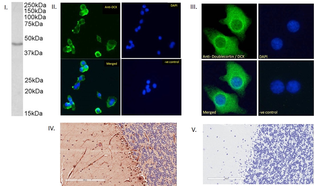
I. (0.01µg/ml) staining of Mouse fetal Brain lysate (35µg protein in RIPA buffer). Detected by chemiluminescence. II. Immunofluorescence analysis of paraformaldehyde fixed HepG2 cells, permeabilized with 0.15% Triton. Primary incubation 1hr (5ug/ml) followed by Alexa Fluor 488 secondary antibody (1ug/ml), showing cytoplasmic staining. The nuclear stain is DAPI (blue). Negative control: Unimmunized goat IgG (10ug/ml) followed by Alexa Fluor 488 secondary antibody (1ug/ml). III. Immunofluorescence analysis of paraformaldehyde fixed KNRK cells, permeabilized with 0.15% Triton. Primary incubation 1hr (10ug/ml) followed by Alexa Fluor 488 secondary antibody (2ug/ml), showing cytoplasmic staining. The nuclear stain is DAPI (blue). Negative control: Unimmunized goat IgG (10ug/ml) followed by Alexa Fluor 488 secondary antibody (2ug/ml). IV. (2µg/ml) staining of paraffin embedded Human Cerebellum. Microwaved antigen retrieval with citrate buffer pH 6, HRP-staining. V. Negative control showing staining of paraffin embedded Human Cerebellum with no primary antibody.
- Heysieattalab, S., Sadeghi, Dynamic structural neuroplasticity during and after epileptogenesis in a pilocarpine rat model of epilepsy, Acta Epileptologica 3, 3 (2021). https://doi.org/10.1186/s42494-020-00037-7 ,0
If you publish research with this product, please let us know so we can cite your paper.


