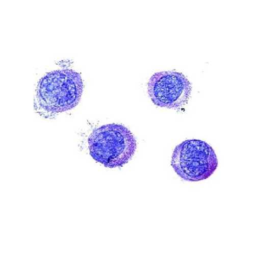
Human Mast Cell Line (LUVA)
Immortalized Human Mast Cell Line (LUVA) that is easily cultured without growth factors.
Highlights:
- Fast growing and increased life span, compared to typical blood-derived CD34+ cells
- Proliferate independent of exogenous stem cell factors
- Contain granules and express high levels of mast cell cytokines, prostaglandins and leukotrienes
Mast cells exist in several types of tissues and play a role in allergy and anaphylaxis as a factor in inflammation response. They are also involved in wound healing and defense against pathogens. These cells express the high-affinity receptors that are specific to IgE, FceRI, on their surface, making IgE molecules cell-surface receptors for an antigen. When bound IgE molecules find a substrate, mast cells release inflammatory mediators like histamine.
From the laboratory of John W. Steinke, PhD, Larry C. Borish, MD, and S. Brandon Early, University of Virginia.
 Part of The Investigator's Annexe program.
Immortalized Human Mast Cell Line (LUVA) that is easily cultured without growth factors.
Highlights:
- Fast growing and increased life span, compared to typical blood-derived CD34+ cells
- Proliferate independent of exogenous stem cell factors
- Contain granules and express high levels of mast cell cytokines, prostaglandins and leukotrienes
Mast cells exist in several types of tissues and play a role in allergy and anaphylaxis as a factor in inflammation response. They are also involved in wound healing and defense against pathogens. These cells express the high-affinity receptors that are specific to IgE, FceRI, on their surface, making IgE molecules cell-surface receptors for an antigen. When bound IgE molecules find a substrate, mast cells release inflammatory mediators like histamine.
From the laboratory of John W. Steinke, PhD, Larry C. Borish, MD, and S. Brandon Early, University of Virginia.
 Part of The Investigator's Annexe program.
This product is for sale to Nonprofit customers only. For profit customers, please Contact Us for more information.
| Product Type: | Cell Line |
| Name: | Human Mast Cell Line (LUVA) |
| Cell Type: | Mast cell |
| Accession ID: | CVCL_5G48 |
| Source: | Blood CD34+ derived |
| Organism: | Human |
| Morphology: | Suspension and adherent |
| Biosafety Level: | BSL1 |
| Subculturing: | Cells can be split 1:2 – 1:20. Cells split 1:10 will be confluent in 3-4 days. |
| Growth Conditions: |
StemPro-34 SFM (500mL), L-glutamine (2mM final), 5mL Pen Strep (10,000 U/mL), 1mL Primocin (50mg). See |
| Cryopreservation: |
90% Complete media, 10% DMSO (See |
| Comments: | The Steinke lab has noted a difference in how the cells are behaving that is different from the original publication. In the article, LUVA cells had high levels of expression of FceRI, however levels of around 5% by cell surface staining are currently being observed by the provider. Higher levels with intracellular staining are seen and very high mRNA expression for FceRI is detected. The reason for this difference is not clear. |
| Storage: | Liquid nitrogen |
| Shipped: | Dry ice |
- Laidlaw, T.M., Steinke, J.W., Tiñana, A.M., Xing, W.,Lam, B.K., Paruchuri, S., Boyce, J.A., and Borish, L. Characterization of a novel human mast cell line that responds to stem cell factor and expresses functional FcεRI. Journal of Allergy and Clinical Immunology 127:815-822.e5 (2011)
- Kirshenbaum AS, Goff JP, Semere T, Foster B, Scott, LM and Metcalfe DD Demonstration that human mast cells arise from a progenitor cell population that is CD34+, c-kit+, and expresses aminopeptidase N (CD13) Blood 1999; 94,7: 2333-2342
- Johnson M, Alsaleh N, Mendoza RP, Persaud I, Bauer AK, Saba L, Brown JM. Genomic and transcriptomic comparison of allergen and silver nanoparticle-induced mast cell degranulation reveals novel non-immunoglobulin E mediated mechanisms. PLoS One. 2018 Mar 22;13(3):e0193499. View Article
If you publish research with this product, please let us know so we can cite your paper.


